Request an Appointment
(703) 719-2040


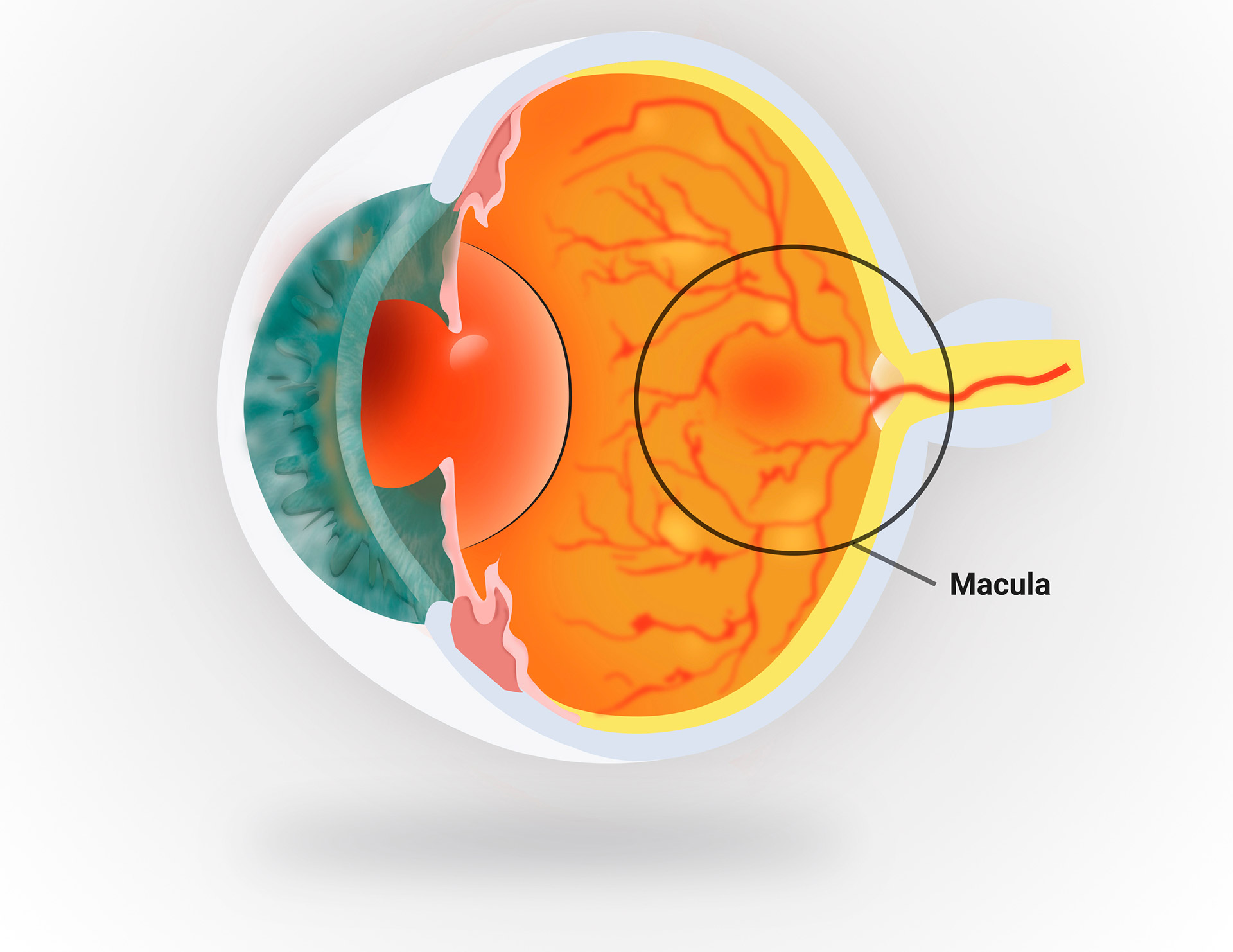
Age-related macular degeneration (AMD) is the most common cause of central vision loss among people 50 years of age and older in developed countries. AMD is a progressive disease that affects the cells in the back part of your eye, which allow you to visualize objects that are straight ahead. With macular degeneration, it becomes increasingly more difficult to perform daily tasks such as reading or writing, driving as well as recognizing faces or colors.
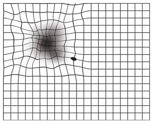
Among the early signs of vision loss from macular degeneration, are shadowy areas in your central vision and unusually fuzzy or distorted vision. While individuals with advanced cases of macular degeneration are considered legally blind as the result of a profound loss of central vision, their peripheral vision, which is less clear than central vision, is retained.
Because most people do not experience vision loss in the early stage of age-related macular degeneration and the progression can be slow and painless, regular dilated retinal examination is vital. Early diagnosis and treatment are the keys to saving vision.
Your ophthalmologist can often detect early signs of macular degeneration before you begin to experience any symptoms and refer you to a retina specialist timely.
Who is at risk for AMD?
You are more likely to develop AMD if you:
Having heart disease is another risk factor for AMD, as is having high cholesterol levels. Caucasians also have an elevated risk for AMD.
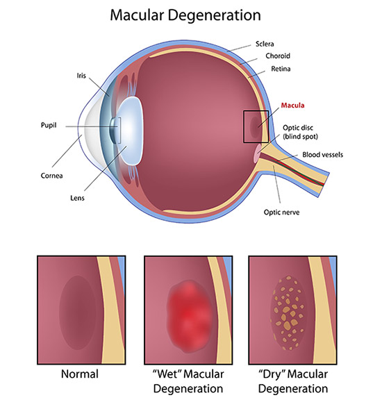
There are two types of AMD: non-neovascular or dry AMD; and neovascular or wet AMD.
Dry AMD
This form is quite common. Dry AMD is when parts of the macula get thinner with age and tiny clumps called drusen appear and grow in the central part of retina. Atrophic areas may also develop. And in the course of time, this process leads to tissue damage and results in central vision loss.
Wet AMD
This form is less common but much more serious. Wet AMD is when new, abnormal blood vessels grow under the retina. These vessels may leak blood or other fluids, causing scarring of the macula. You lose vision faster with wet AMD than with dry AMD.
Many people don’t realize they have AMD until their vision is very blurry. Therefore, it is important to have regular visits to an eye doctor. Your doctor can look for early signs of AMD before you have any vision problems and refer you to a retina specialist timely.
If you have dry AMD, proper diet with a special formula of vitamins and minerals called AREDS II, based on the Age-Related Eye Disease Study II, can help slow down progression of the disease and preserve your vision.
If you have wet AMD, ocular injections of vision saving medicines can not only preserve your vision, but may reverse vision loss significantly. Your retina specialist at Retina and Uveitis Center is uniquely trained and qualified to give you these injections and monitor your progress.
Although presently there is no known cure for macular degeneration, our office can help you learn about the treatment options that may prevent or slow its progression.
AMD causes your vision to change over time. You may not notice these changes when they happen. But you need to catch vision changes as soon as possible. Treating them early can help slow or stop further vision loss.
You should use an Amsler grid regularly to monitor your vision.
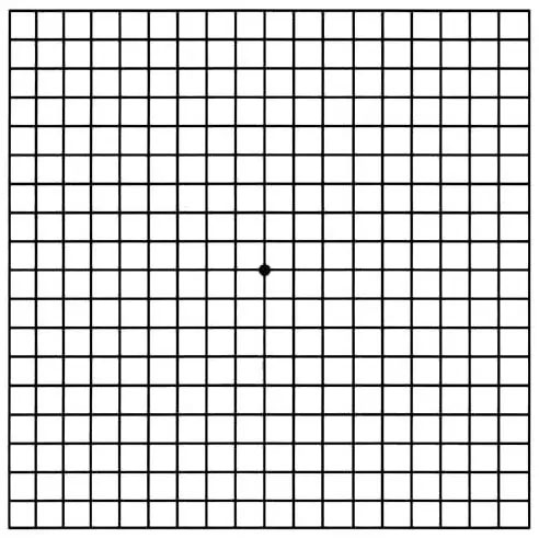
Here is how to use the Amsler grid:
Call your retina specialist right away, if you notice that any lines or parts of the grid look wavy, blurry, or dim.
This is what an Amsler grid might look like with blurry, wavy lines, or dim areas.