Side by side retinal fundus photographs of both eyes showing optic discs, blood vessels and maculas for retina examination
Hand pointing a pen at retinal scans on a monitor while a masked patient sits at an eye exam machine

EXPERIENCED CARE YOU CAN TRUST

Welcome to Retina and Uveitis Center!

We offer medical and surgical specialty services to patients and referring doctors from a wide regional referral area for evaluation and management of vitreoretinal and ocular inflammatory diseases. Our mission is to provide state-of-the art vision care tailored to the needs of the patients and the referring doctors in a timely and compassionate manner.



2025 Washingtonian Top Doctors plaque awarded to Muge R. Kesen, MD, for ophthalmology, selected by peers Washingtonian Top Doctor 2026 award badge in blue lettering on a white background

Specializing in the Evaluation and Management of Retinal Diseases

Retina and Uveitis Center | Retinal Artery Occlusion, Pattern Dystrophy and Dislocated IOL
Macular Degeneration
A progressive disease that affects the cells in the back part of your eye, which allows you to visualize objects that are straight ahead. Over time it damages your capacity for sharp, central vision.
Fundus photograph of a healthy retina showing the optic disc, macula, and branching blood vessels used in retinal exams
Diabetic Retinopathy
The most common cause of vision loss among individuals with diabetes is diabetic retinopathy, which is a consequence of damage to the tiny blood vessels in the back of your eyes.
Retina fundus photo showing yellow deposits around the macula, as seen in macular degeneration and other macular disease
Retinal Vein Occlusion
RVO occurs when the blood flow heading back to the heart gets blocked. With no means for the blood to drain, hemorrhages (bleeding) and leakage from the blocked blood vessels can ensue.
Abstract blue background with overlapping diagonal rectangles in varying shades creating a layered geometric pattern
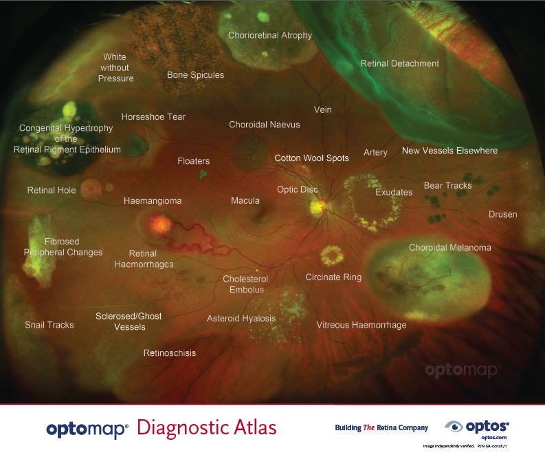
Optos widefield retinal scan labeled with conditions like retinal detachment, horseshoe tear, cotton wool spots and drusen

Optos® - ultra-wide field retinal imaging

Optos' devices have the unique ability of producting an ultra-widefield, high resolution digital image of approximately 82% and 200 degree of the retina all in a single image.

Get to know our Doctor

Muge R. Kesen, MD

Dr. Kesen is a clinical-researcher with over 10 years of experience both in academic and private practice settings with exceptional commitment to patient care and dedication to clinical research. She specializes in vitreoretinal and inflammatory diseases of the eye. She is a member of the American Society of Retina Specialists (ASRS) and the American Academy of Ophthalmology (AAO).

Learn More

Dr. Muge R. Kesen, MD smiling in a black blazer, retina and uveitis specialist at the practice
Side by side retinal fundus photographs of both eyes showing optic discs, blood vessels and maculas for retina examination

THERE’S NO TIME LIKE THE PRESENT

SEEK TREATMENT TODAY

Advanced Technology

Precise and Quality Care

American Society of Retina Specialists logo with a blue circular emblem beside the letters ASRS
Retina and Uveitis Center | Macular Edema, Macular Degeneration and Diabetic Retinopathy
American Academy of Ophthalmology logo with a purple starburst circle and the tagline Protecting Sight. Empowering Lives.
2023 Northern Virginia Magazine Top Doctors award badge recognizing retina care specialists
American Academy of Ophthalmology EyeSmart banner offering expert information about eye health for patients
Arlington Magazine Top Doctor 2024 award badge on a blue background with white and dark gray text
Side by side retinal fundus photographs of both eyes showing optic discs, blood vessels and maculas for retina examination