Request an Appointment
(703) 719-2040


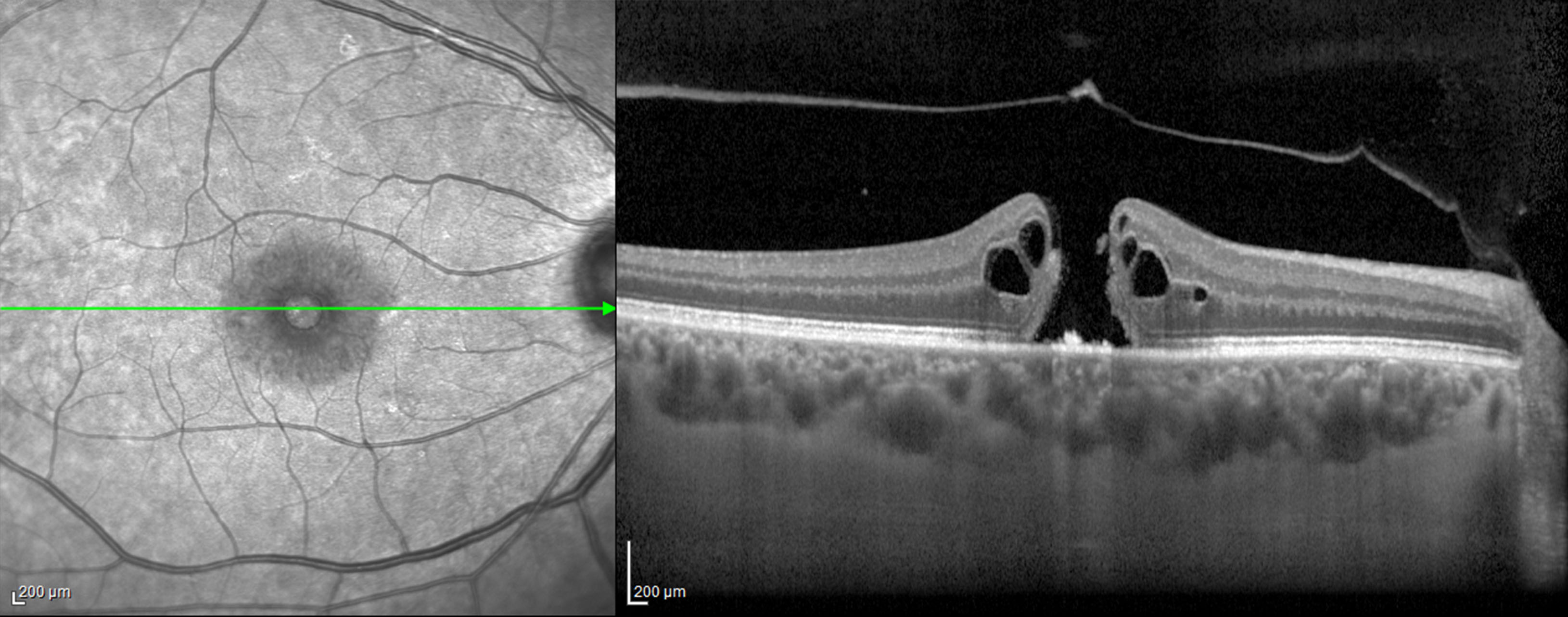
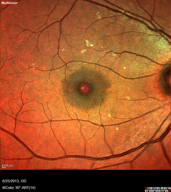
The macula is a small area in the center of the retina where light is sharply focused to produce the detailed color vision needed for tasks such as reading and driving. Although it’s relatively rare, some people develop a full-thickness defect in the macula, and the condition is referred to as macular hole. Because the macula plays a significant role in your central vision, a macular hole can affect your ability to see straight ahead, read or view the fine details.
Age is the primary risk factor for the development of macular hole, but additional risk factors include:
It’s also noteworthy that if a macular hole develops in one eye, there is also risk of developing one in the other eye. And for unknown reasons, women are at greater risk of developing this disorder than men.
While the general symptoms include gradual decline and blurriness in central vision, a wavy-looking appearance of straight lines, and trouble reading the fine print, you may also experience gray or black spots in your central vision. The severity of your symptoms depends upon the macular hole’s size, location, and its developmental stage.
Sometimes a tiny macular hole can close on its own. However, in cases where surgery is indicated, vitrectomy is the most common procedure for treatment of macular holes. In this short outpatient surgical procedure, the vitreous gel is removed to stop it from pulling on the retina, and a gas bubble is placed in the eye to gently hold the edges of the macular hole closed until it heals. The patient may be asked to maintain a face-down position for several days, and in some cases up to 2 weeks, depending on the characteristics of the macular hole. This will allow the bubble to gradually dissolve and be replaced by natural eye fluids.
As with all eye conditions, it’s essential to be ever mindful of your vision health. Our office keeps a close watch on the health of your eyes and the quality of your vision.