Request an Appointment
(703) 719-2040

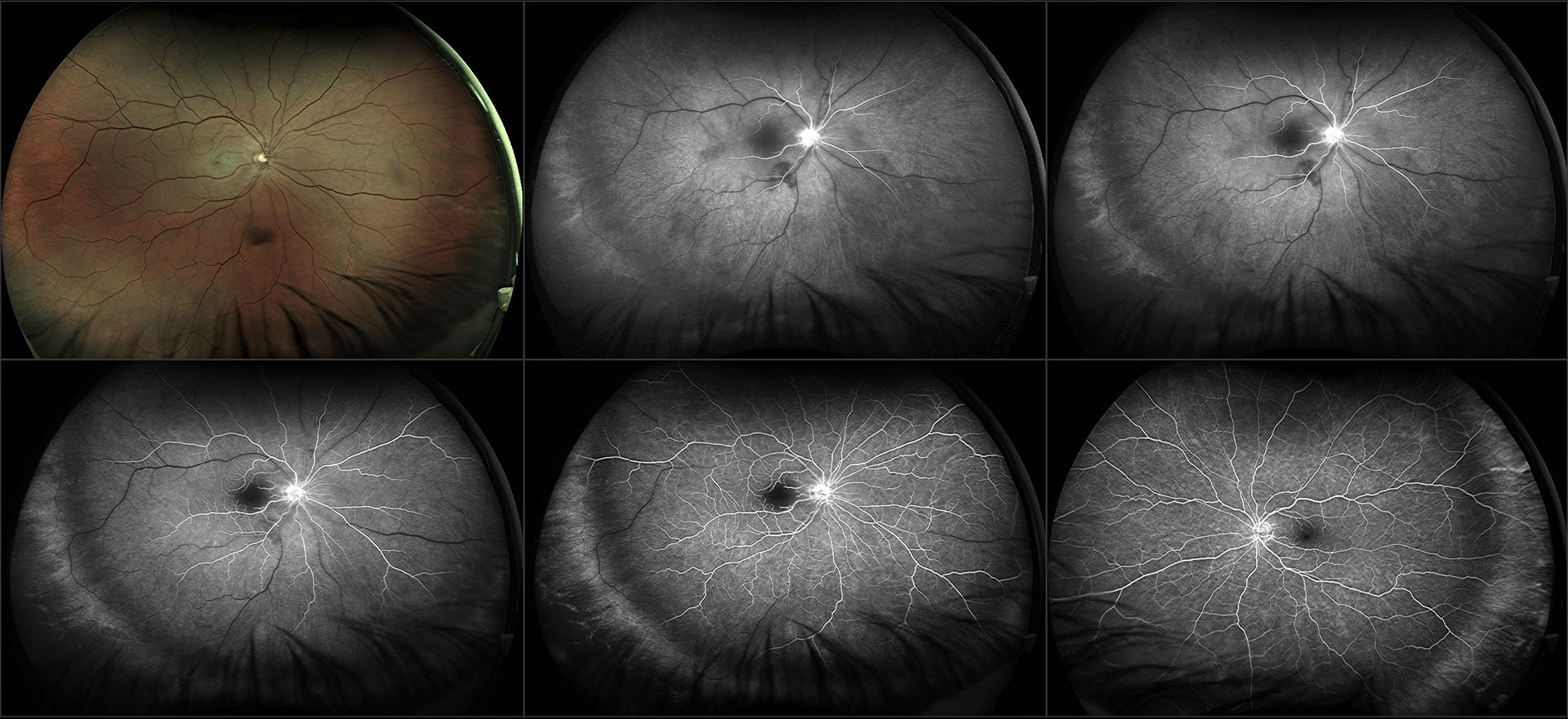
The task of bringing oxygen and nutrients to the retina primarily falls to the central retinal artery (CRA) and its branches. When this artery or its branches get partially blocked or occluded due to a retinal embolism (a small piece of cholesterol that blocks blood flow), thrombus (blood clot), atherosclerosis, or arteritis, it can result in varying degrees of vision loss. The retinal artery occlusion may be transient and last for only a few seconds or minutes if the blockage breaks up and restores blood flow to the retina, or it may be permanent.
Retinal artery occlusion is usually associated with sudden painless loss of vision in one eye. The area of the retina affected by the blocked vessels determines the area and extent of visual loss.
Common risk factors include:
Most retinal artery occlusion patients are in their 60s and are more commonly men than women.
Unfortunately, there is no clinically proven treatment for CRAO.
Persons with an acute onset CRAO or BRAO are referred promptly to the emergency room or their primary care doctor to be evaluated for stroke risk. An important aspect of managing retinal artery occlusion is for your primary doctor to identify and manage risk factors that may lead to other vascular conditions. The risk factors for CRAO are the same atherosclerotic risk factors as for stroke and heart disease.
Formation of new blood vessels of the retina or iris that are prone to bleed is a rare complication seen after a CRAO or BRAO. Growth of these vessels can further decrease vision by causing vitreous hemorrhage and glaucoma. Intraocular injections and/or laser photocoagulation therapy may need to be used in such cases.
