Request an Appointment
(703) 719-2040


The retina's role often gets compared to the function film plays in a conventional camera. With millions of photoreceptors known as rods and cones, the retina translates focused light into the full-color visual information that gets sent to the brain.
In a healthy retina, this intricate process operates seamlessly. However, when a retinal disorder affects this area in the eye, it can cause vision impairment, low vision, and even blindness.
A detached retina occurs when the light-sensitive tissue layer at the back of the eye gets pulled away from the underlying supporting tissue—thereby separating the retina from its blood and oxygen supply. In some cases, a retinal detachment happens after a retinal tear when vitreous fluid seeping through the opening detaches the retina from the tissue underneath.
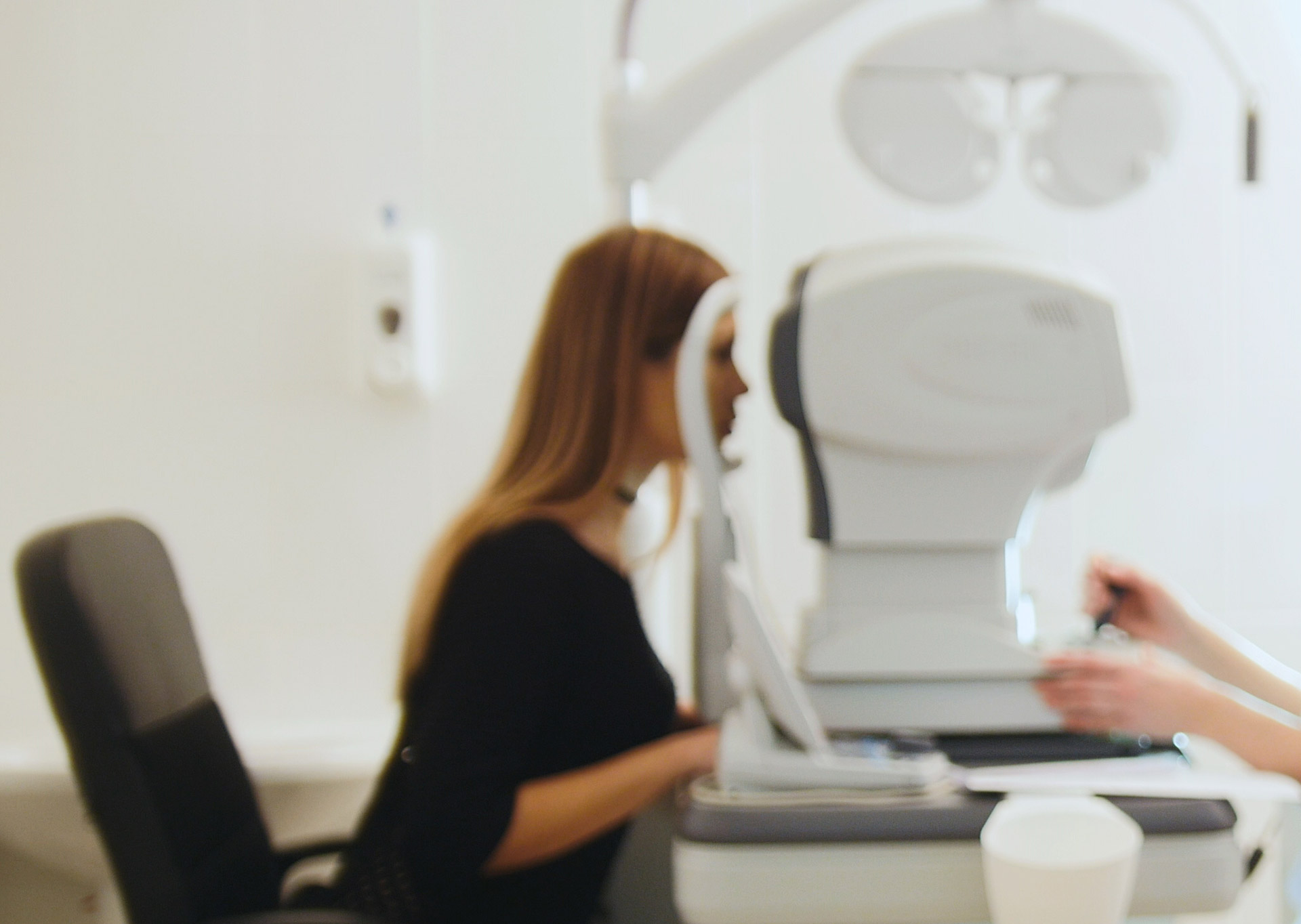
A dilated eye exam gives the eye doctor a view of the back of the eye and an opportunity to check for evidence of retinal detachment, holes, or tears. Also, when indicated, ultrasound and optical coherence tomography (OCT) can offer more detailed information, especially when the retina is obscured.
Because the photoreceptors begin to deteriorate quickly when a retinal detachment occurs, it's essential to get a prompt evaluation and care. A retinal detachment can worsen rapidly and lead to permanent vision loss or even blindness. Plus, the longer the retina has been detached, there's less likelihood vision can get restored.
It's essential to be aware of any sudden onset vision changes, including:
The goal of repairing a retinal detachment is to place the light-sensitive tissue back into its normal position, restore vision as possible, and prevent permanent vision loss. While some repairs may require more than one surgery, statistics demonstrate that more than 9 out of 10 detachments can get repaired. However, it's too late to restore normal vision when a detachment occurs in the macula, which is responsible for central vision. In this case, surgery aims to prevent further vision loss and total blindness.
Since a retinal tear puts you at increased risk of developing a detached retina, the tear should get repaired with laser photocoagulation or a cryopexy procedure.
Repairing a detached retina involves one or a combination of the following procedures:
It's essential to keep in mind that the type of surgery required depends on the extent of the detachment and where it is located. It's not uncommon to undergo a combination of procedures to achieve optimal success. Following retinal detachment repair, it can take several months to see improvements.